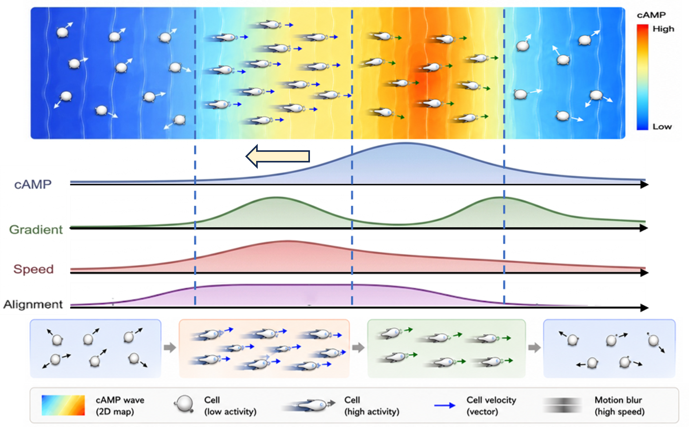

New imaging method reveals that individual cells behave like surfers — paddling toward an incoming
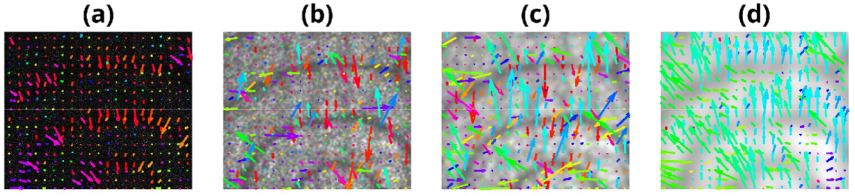
chemical wave, riding its crest, then drifting until the next wave arrives
Imagine a surfer paddling out to sea — not chasing a single wave, but timing their movement to a rhythm. As the next swell approaches, they paddle hard toward the crest. They ride it forward, then, once it passes, they float, drifting aimlessly until the next wave arrives. This isn’t just a metaphor — it’s how Dictyostelium discoideum, a social amoeba, survives starvation.
When food runs out, thousands of individual cells stop roaming and begin emitting a chemical signal — cyclic AMP (cAMP) — in rhythmic, spiral waves across the colony. These waves act like invisible surfboards, guiding cells to move together, form a multicellular slug, and eventually produce spores to ensure the species survives.
But how do individual cells “know” when to move — and in which direction?
A new study published in Scientific Reports reveals the answer: they surf. Using a clever twist on imaging technology, researchers from different universities discovered that individual amoeba cells don’t just react to cAMP waves — they anticipate them.
By applying Gaussian blurring to time-lapse fluorescence images, the team uncovered a hidden layer of biological rhythm. Using particle image velocimetry (PIV), a method borrowed from fluid dynamics, they extracted Eulerian velocity vector fields from the raw image sequences.
At fine scales, unblurred images revealed the actual movement of individual cells: directed, phase-locked motion toward the approaching cAMP wave. But as the images were progressively blurred, the individual cell trajectories faded into the background — and a smooth, sweeping cAMP wave emerged as the dominant signal in the PIV vector field.
To understand when cells respond, the researchers used wavelet-based multiscale phase analysis. This technique identifies the precise phase of the cAMP wave — from rising to peak to falling — even in noisy, non-stationary signals. It works by decomposing the cAMP intensity over time into multiple frequency components, filtering out high-frequency noise, and extracting the true oscillatory rhythm.
The results were striking: cells consistently moved toward the approaching wave crest, but only during specific phases — particularly when the cAMP concentration was rising. After the wave passed, their motion became randomized, with no clear direction.
This revealed a phase-dependent surfing behavior: cells actively orient and accelerate toward the oncoming wave, then rest in the troughs until the next cycle begins.
This collective surfing behavior — where cells engage purposefully with an oncoming signal, then rest until the next wave arrives — offers one of the first systematic, quantitative windows into how independent cells transition into a synchronized collective.
The method is revolutionary. It requires no individual cell tracking, which is often impossible in dense, aggregating populations. Instead, by analyzing motion at different spatial scales using PIV and phase information from wavelet analysis, the team extracted the true rhythm of collective behavior from the same footage that once only appeared to show individual motion.
The approach could transform how we study wave-mediated collective behaviors in biology — from neutrophil swarms during immune responses to wound healing and even cardiac tissue dynamics.
Publication:
Sattari, S. et al. “Collective surfing of single cells on a chemo-attractant wave using multiscale Eulerian velocity vector field.” Scientific Reports (2026). DOI: 10.1038/s41598-026-61774-2