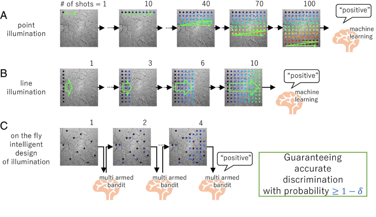
Many diseases are difficult to identify based solely on information from stained images of cells and tissues. An analytical technique called Raman spectroscopy can comprehensively provide chemical information about the molecules that exist in a sample without damaging cells or tissues, thus making it a promising tool for medical diagnosis. However, Raman spectroscopic measurements are time-consuming, limiting their practical use. Now, a multi-institute team led by ICReDD principal investigator Professor Tamiki Komatsuzaki has developed a system that uses artificial intelligence to enable accurate Raman spectroscopy measurement that is hundreds to thousands of times faster than conventional methods.
The team developed an on-the-fly system that uses a reinforcement learning algorithm to optimize which locations on the sample are measured, rather than measuring every single point. During sample measurement, a feedback loop based on a “multi-armed bandit” algorithm analyzes the data obtained thus far in the measurement, and selects the subsequent measurement locations so as to efficiently and rapidly measure areas with a high probability of containing abnormalities.
Researchers used a mix of two different polymer beads as a model case to mimic malignant abnormalities and normal tissue. For this proof-of-concept case, the on-the-fly Raman spectroscopy method was hundreds to thousands of times faster at discriminating between samples with and without abnormalities compared to conventional, raster-scanning Raman spectroscopy. Additionally, users can preset the required accuracy, making the system adaptable to various user needs.
This leap in the rapid measurement of Raman spectroscopy is expected to lead to various biological applications including diagnoses that are difficult using staining techniques, determination of the severity of breast cancer, assessment of the maturity of stem cells, and evaluation of drug responses in liver and cancer cells. Additionally, this method could potentially be used for anomaly detection in semiconductors and inspection of microplastics.