The following text is adapted from a joint press release led by Nagoya University.
Key points
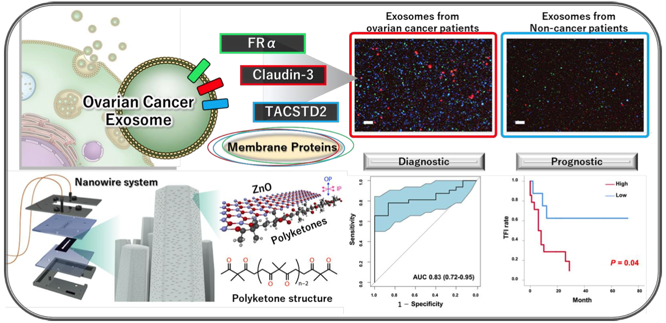
・Revealed detailed protein information on ovarian cancer EVs and their diversity
・Development of polyketone-coated nanowires for simple EV isolation
・Discovery of ovarian cancer specific membrane proteins and their utility
・Expected to be a new EV biomarker strategy to improve ovarian cancer treatment
Research Summary
Extracellular vesicles (EVs), including exosomes, are present in all human body fluids and have attracted attention as an essential tool for intercellular communication. Membrane proteins on the surface of EVs are necessary to detect specific EVs. They can be used as biomarkers on their own. However, specific EV membrane proteins in ovarian cancer are not known. Ovarian cancer is a female genital malignancy with a poor prognosis. It is one of the leading causes of cancer death among women worldwide. Ovarian cancer is one of the most challenging cancers to detect in its early stages, and the development of a highly accurate and sensitive biomarker is urgently needed. This study identified ovarian cancer EV-associated membrane proteins, FRα, Claudin-3, and TACSTD2, through detailed proteomic analysis in ovarian cancer-derived EVs. Nanowire systems are one of the methods for capturing EVs, and this study succeeded in attaching polyketone chains to ZnO nanowires, which enabled capturing EVs with higher purity. Using this new method, we have developed a novel detection method using EVs in ovarian cancer patients. These findings hold promise as a new biomarker strategy for ovarian cancer.