A team of researchers from Hokkaido University’s Faculty of Medicine and ICReDD developed a new method that can spatially detect Rac1 and Cdc42 regulatory factors, which are associated with cancer tumor invasion. The ability to detect these factors level of activation and their positions reliably could aid greatly in predicting cancer behavior in individual patients, enabling more personalized medicine.
Traditional methods to detect activation involved heating the sample and the use of hazardous isotopes, which led to a loss of spatial information. The technique reported here preserves spatial information by combining a special probe molecule with the use of electrical micro-agitation to increase binding of the probe with the proteins of interest. In the study, 29 of 33 human colon cancer tissue samples showed higher activity of these Rac1 and Cdc42 in the tumor area compared to normal mucosa, with a striking amount of activity at the invasive front of the tumor. Use of this technique could help to better understand how the activation of these factors relates to tumor growth and spread.
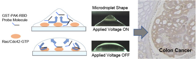 Schematic (left) of the changes in the microdroplet as the voltage is switched on and off. The electrical micro-agitations stirs the probe solution and increases the opportunity for binding between the probe and Rac/Cdc42-GTP. This enables spatially resolved detection of Rac/Cdc42 activity in colon cancer tissue samples (right).